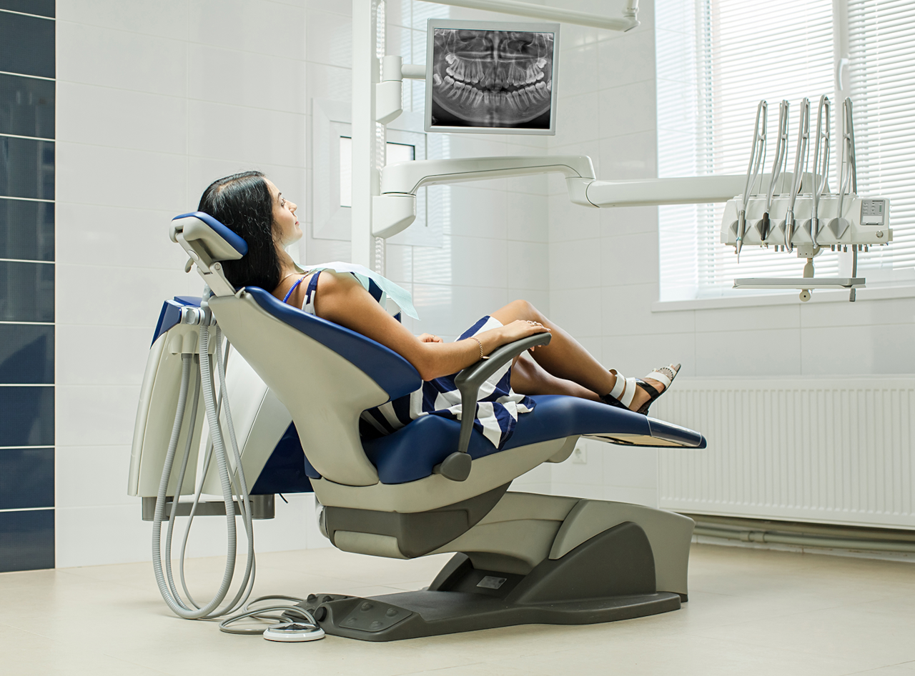
A dental examination can reveal a great deal about your smile, but some important problems develop in places that cannot be seen by looking inside the mouth alone. Cavities can form between teeth, infections can develop around tooth roots, bone levels can change beneath the gums, and impacted teeth can remain hidden below the surface. Dental X-rays give your dentist another way to evaluate these areas before a problem becomes obvious from appearance or symptoms.
At AB Dental and Oral Surgery, preventive dentistry in San Antonio includes comprehensive examinations and diagnostic tools that help the dental team evaluate your oral health more completely. Digital dental X-rays can provide detailed images that support diagnosis, treatment planning, and ongoing monitoring while using modern imaging technology designed for efficient dental care.
Patients often have questions about whether X-rays are necessary at every visit, how much radiation they involve, and why different types of dental images are sometimes recommended. The answer is that dental X-rays should be based on individual needs rather than a one-size-fits-all schedule. Your age, cavity history, gum health, symptoms, previous dental treatment, and current oral condition can all influence which images are useful and when they should be taken.
What Are Dental X-Rays?
Dental X-rays are diagnostic images that allow dentists to view structures that are not fully visible during a routine visual examination. Depending on the type of image, an X-ray can show the crowns of teeth, roots, surrounding bone, developing teeth, jaw structures, and other areas that may need evaluation.
Digital dental X-rays use electronic sensors or other digital imaging systems to capture information that can be displayed on a computer screen. This allows the dental team to enlarge, adjust, and review the image while discussing findings with the patient.
X-rays do not replace a clinical examination. Instead, they add another layer of information. A dentist still needs to look at the teeth and gums, ask about symptoms, evaluate the bite, and perform other tests when appropriate.
Why Can a Dentist Need X-Rays if My Teeth Feel Fine?
Many dental problems do not cause symptoms in their earliest stages. A small cavity between two teeth may not hurt. Bone loss from periodontal disease may develop gradually. A tooth with a developing problem around the root may initially feel normal.
If dentists relied only on pain, some conditions could become significantly more advanced before they were identified. Dental X-rays can help reveal changes before they become easy to see or feel.
This does not mean every patient needs every possible X-ray at every appointment. The purpose of imaging is to answer specific diagnostic questions and supplement what the dentist learns from the clinical examination.
What Can Dental X-Rays Show?
The information provided by a dental X-ray depends on the type of image and the area being examined. Dental images may help a dentist evaluate:
- Cavities between neighboring teeth
- Decay beneath or around existing restorations
- Tooth roots and the surrounding bone
- Changes associated with dental infection
- Bone levels around teeth affected by gum disease
- Impacted or unerupted teeth
- Wisdom tooth position
- Developing permanent teeth in younger patients
- Previous root canal treatment
- Areas being considered for dental implants
- Jaw structures and selected abnormalities
- The relationship between teeth and nearby anatomy
No single dental X-ray provides every type of information. Different views are used because each is designed to answer a different clinical question.
What Are Bitewing X-Rays?
Bitewing X-rays are commonly used to view the upper and lower back teeth in the same region. They can be particularly useful for evaluating areas between teeth where cavities may be difficult to see directly.
These images can also help dentists assess existing fillings and crowns, look for changes near restoration margins, and evaluate portions of the supporting bone.
Bitewings are called bitewings because the patient traditionally bites on a small tab or positioning device while the image is captured.
What Are Periapical X-Rays?
A periapical X-ray focuses on one tooth or a small group of teeth and usually shows the entire tooth from the crown through the root and into the surrounding bone.
This type of image may be useful when a patient has tooth pain, a suspected infection, a history of root canal therapy, trauma, or another concern involving the root or surrounding structures.
Because the full root is visible, periapical imaging can provide information that a bitewing image may not capture.
What Is a Panoramic Dental X-Ray?
A panoramic image provides a broad view of the jaws, teeth, and surrounding structures. Instead of placing a small sensor next to individual teeth, the imaging equipment moves around the head to create a wider picture.
Panoramic X-rays can be helpful when evaluating wisdom teeth, tooth development, multiple missing teeth, jaw relationships, or other situations where a broad overview is useful.
A panoramic image does not necessarily replace smaller intraoral X-rays. Fine details needed to identify certain cavities or localized problems may require another type of image.
What Are Digital Dental X-Rays?
Digital dental X-rays use modern electronic imaging technology rather than relying solely on traditional film. The resulting images can be displayed quickly and reviewed on a computer.
This digital format provides several practical advantages. The dentist can enlarge areas for closer evaluation, compare images over time, and show the patient what is being examined.
Digital images can also be stored as part of the dental record, making them useful for monitoring changes at future appointments.
Benefits of Digital Dental X-Rays
Digital dental X-rays can provide valuable information that complements a visual dental examination. When imaging is recommended according to the patient’s needs, it can help the dental team identify, monitor, and plan treatment for conditions that may otherwise remain hidden.
- Detects hidden dental problems: X-rays can reveal certain cavities, infections, impacted teeth, and bone changes that may not be visible during a visual examination.
- Supports earlier diagnosis: Finding a problem before it becomes larger may allow more treatment options to remain available.
- Helps evaluate tooth roots: Periapical images can show roots and surrounding bone that cannot be seen by looking at the crown of the tooth.
- Assists with periodontal evaluation: Dental images can help show bone levels around teeth when gum disease is a concern.
- Improves treatment planning: Imaging can provide information needed when planning extractions, root canal therapy, implants, crowns, and other procedures.
- Allows comparison over time: Previous digital images can be compared with newer ones to monitor changes.
- Provides visual patient education: Digital images can be enlarged on a screen so patients can better understand areas being discussed.
- Supports individualized preventive care: Imaging frequency can be adapted to cavity history, symptoms, gum health, age, and other risk factors.
Do You Need Dental X-Rays at Every Visit?
Not necessarily. Dental X-ray frequency should be based on individual clinical needs rather than automatically taking the same images at every appointment.
A patient with a history of frequent cavities may need certain images more often than someone who has consistently healthy teeth and a low risk of decay. A patient with periodontal disease may need imaging that helps monitor bone levels. Someone with sudden tooth pain may need a targeted image even if routine X-rays were taken relatively recently.
Children and teenagers may also need imaging for reasons that differ from those of adults because their teeth and jaws are still developing.
Your dentist can determine which images are justified based on your examination, dental history, previous X-rays, and current concerns.
How Often Should Adults Have Dental X-Rays?
There is no single interval that is correct for every adult. The appropriate schedule can depend on cavity risk, gum disease, existing restorations, symptoms, oral hygiene, dry mouth, previous dental treatment, and other factors.
An adult with a low risk of cavities and no active dental concerns may not need bitewing images as frequently as a patient who develops new decay regularly.
Likewise, a patient with a new toothache may need an image outside the usual preventive schedule because the dentist is investigating a specific symptom.
Rather than focusing only on how long it has been since the last X-ray, the more useful question is what information the dentist needs now to evaluate your oral health.
Why Might Children Need Dental X-Rays?
Children’s mouths change quickly as primary teeth are lost and permanent teeth develop. Dental images may help monitor tooth eruption, identify cavities between teeth, evaluate dental development, and assess areas that cannot be seen clinically.
Children who have a higher cavity risk may require a different imaging schedule from children with consistently healthy teeth.
The decision should consider the child’s age, dental development, history of decay, spacing between teeth, symptoms, and previous images.
Are Dental X-Rays Safe?
Dental professionals use X-rays when the expected diagnostic information is considered useful for patient care. Modern digital dental imaging is designed to capture the needed information efficiently, and practices use protective measures and appropriate imaging protocols.
Radiation exposure is one reason dentists do not take unnecessary images simply out of routine habit. Instead, the type and frequency of imaging should match the clinical need.
If you have concerns about radiation exposure, discuss them with your dentist. The dental team can explain why a particular image is being recommended and what information it is expected to provide.
How Much Radiation Comes From a Dental X-Ray?
Radiation dose varies according to the type of dental image, the equipment being used, and the imaging technique. A small intraoral image and a broader panoramic image do not involve exactly the same exposure.
For patients, the most important point is that dental imaging should be justified by diagnostic need and performed using modern techniques and appropriate settings.
Instead of avoiding necessary imaging because radiation exists, patients can ask why an image is needed and how it will influence diagnosis or treatment planning.
Why Does the Dental Team Ask About Pregnancy?
Pregnancy is an important part of your medical history, so you should tell the dental team if you are pregnant or think you may be pregnant.
Dental care sometimes remains necessary during pregnancy, particularly when pain, infection, or another urgent problem needs diagnosis and treatment. Your dentist can determine whether imaging is needed and what precautions are appropriate for the situation.
Do not delay reporting severe dental pain or swelling because of pregnancy concerns. Instead, provide accurate medical information so your dental and medical providers can make informed decisions about your care.
Can Dental X-Rays Find Cavities Before They Hurt?
Yes. Certain cavities may be visible on dental X-rays before they produce symptoms.
Decay between two neighboring teeth is a common example. The contact area may prevent the dentist from seeing the entire surface directly during a visual examination.
Bitewing images can help show changes in these areas. Identifying decay earlier can be useful because a smaller area may require less extensive treatment than a cavity that has progressed deeper into the tooth.
Can Dental X-Rays Show an Infection?
Dental X-rays can sometimes reveal changes in the bone around a tooth root that are associated with infection or inflammation. However, imaging is only one part of diagnosis.
A dentist may also ask about pain, swelling, temperature sensitivity, biting discomfort, previous dental treatment, and other symptoms.
The tooth may be tested for tenderness, nerve response, mobility, or bite pressure. Several pieces of information may be combined before treatment is recommended.
Not every infection looks identical on an X-ray, particularly in its earliest stages. Therefore, a normal-looking image does not automatically explain every symptom.
Can Dental X-Rays Show Gum Disease?
Dental X-rays can help show the height and pattern of bone around teeth. This information is important when evaluating periodontal disease because advanced gum disease can damage the bone supporting the teeth.
However, X-rays cannot replace periodontal measurements and a clinical gum examination.
Your dental team may measure the spaces between the teeth and gums, look for bleeding or inflammation, check for tartar buildup, evaluate recession, and use imaging to assess supporting bone.
Together, these findings provide a more complete picture of periodontal health.
Can an X-Ray Show a Cracked Tooth?
Sometimes, but not always. Cracks can be difficult to identify on standard dental X-rays because their visibility depends on their direction, size, location, and the angle of the image.
This is why dentists often use several diagnostic methods when a cracked tooth is suspected.
The dentist may evaluate biting pain, use magnification, inspect the tooth visually, check existing restorations, test the bite, and review dental images.
A patient should not assume that a normal X-ray automatically means a crack is impossible.
Why Are X-Rays Important Before Certain Tooth Extractions?
When a tooth needs to be removed, dental imaging can help show root shape, root position, surrounding bone, nearby teeth, and other anatomical considerations.
This information can be especially important for teeth with complex roots or teeth that have not fully erupted.
Wisdom teeth are one example. A broader image may help the dentist evaluate their position and relationship to nearby structures before planning treatment.
Why Are Dental X-Rays Used for Root Canal Treatment?
Root canal therapy involves the internal spaces of a tooth, including areas extending through the roots. Dental imaging can provide information about root anatomy, surrounding bone, previous treatment, and possible signs of infection.
Images may be used at different stages depending on the case.
Because the internal anatomy is not visible from the outside of the tooth, diagnostic imaging plays an important role in understanding what is happening beneath the surface.
Why Are X-Rays Important for Dental Implants?
Dental implants are placed within the jawbone, so evaluating bone and nearby anatomy is an essential part of implant planning.
The imaging required for implant treatment may differ from standard preventive dental X-rays. In certain cases, more advanced three-dimensional imaging may be recommended to provide additional information about bone dimensions and anatomical structures.
The appropriate imaging depends on the implant location and complexity of treatment.
What Is a CBCT Scan?
Cone beam computed tomography, often shortened to CBCT, is a type of dental imaging that can create three-dimensional views of teeth, bone, and surrounding structures.
A CBCT scan provides different information from a routine two-dimensional dental X-ray. It may be used when more detailed anatomical information is needed for certain surgical, implant, endodontic, or other complex dental situations.
Not every dental patient needs a CBCT scan. Three-dimensional imaging should be selected when the additional information is relevant to diagnosis or treatment planning.
Can Dental X-Rays Show Oral Cancer?
Dental imaging can sometimes reveal unusual changes in bone or other structures, but X-rays are not a substitute for an oral cancer screening.
An oral cancer examination includes looking at and feeling the soft tissues of the lips, tongue, cheeks, gums, palate, and other accessible areas.
If an abnormal area is found, additional evaluation or testing may be necessary depending on the location and characteristics of the finding.
Do Dentists Keep Previous X-Rays?
Dental images are generally maintained as part of the patient’s dental record according to applicable recordkeeping requirements and practice procedures.
Previous images can be valuable because they allow the dental team to compare changes over time.
For example, a dentist may compare current bone levels, restoration margins, or tooth conditions with previous images to see whether a change is new or has remained stable.
Should You Send Old X-Rays to a New Dentist?
If you are changing dental offices, recent dental records and X-rays may be useful to the new practice. Previous images can provide history and may help prevent unnecessary duplication when the existing images are current and diagnostically useful.
However, a new dentist may still recommend updated images if the previous set does not show the areas currently being evaluated, is no longer current enough for your risk level, or if new symptoms have developed.
The decision should be based on whether the available images answer the clinical questions at the new appointment.
What Should You Tell Your Dentist Before X-Rays?
Provide an accurate medical and dental history before treatment. Important information may include pregnancy, recent dental X-rays at another office, new symptoms, previous dental procedures, and health conditions that could influence your dental care.
If you have had imaging taken recently elsewhere, mention it. The dental team may be able to request those records and determine whether additional images are needed.
You should also describe exactly where you are experiencing pain or sensitivity, because this can help the dentist select the most useful view.
Why Can X-Rays Be Necessary Even After a Recent Cleaning?
A professional cleaning removes plaque and tartar, but it does not provide an image of internal tooth structure or bone.
A patient can have very clean teeth and still develop a cavity between teeth, an infection near a root, an impacted tooth, or another hidden concern.
Cleaning and diagnostic imaging therefore serve different purposes. One focuses on removing deposits from accessible tooth surfaces, while the other provides information about structures that cannot be fully examined visually.
Can You Refuse Dental X-Rays?
Patients can ask questions about any recommended dental procedure, including imaging. If you are uncertain about an X-ray, ask your dentist why it is being recommended, what area it will show, and how the information may affect your treatment.
However, there are situations where a dentist may not be able to diagnose or safely plan treatment without appropriate imaging.
For example, attempting certain procedures without understanding root position, bone condition, or hidden decay could leave important information unknown.
A clear conversation about the purpose of imaging can help you make an informed decision.
How Do Digital X-Rays Help Patients Understand Their Dental Health?
Digital imaging is useful not only for the dentist, but also for patient education.
An image can often be displayed on a larger screen so your dentist can point out the area being discussed. You may be able to see the location of a cavity, a restoration, an impacted tooth, or changes in supporting bone.
This can make treatment recommendations easier to understand because the conversation is connected to a visual representation of your own mouth.
Patients should still feel comfortable asking questions. An image can provide useful information, but the dentist should explain what the finding means and what treatment options are available.
How Can You Reduce the Need for Problem-Focused Dental X-Rays?
You cannot eliminate every reason dental imaging might be needed. However, maintaining good oral health can reduce the number of unexpected problems that require urgent diagnostic evaluation.
- Brush twice daily with fluoride toothpaste.
- Clean between your teeth every day.
- Keep routine dental examinations.
- Attend professional cleanings as recommended.
- Limit frequent sugary snacks and beverages.
- Address dry mouth if it becomes a persistent problem.
- Report tooth sensitivity or biting pain early.
- Have damaged fillings and crowns evaluated promptly.
- Seek care for swelling or signs of infection.
- Wear appropriate mouth protection during activities with a risk of dental injury.
These habits cannot replace diagnostic imaging when it is needed, but they can support healthier teeth and help identify problems before they become dental emergencies.
Frequently Asked Questions About Dental X-Rays
Do I need dental X-rays at every dental appointment?
Not necessarily. Imaging frequency should be based on your cavity risk, gum health, symptoms, dental history, age, previous images, and the information your dentist needs for diagnosis or treatment planning.
Can dental X-rays detect cavities?
Yes. Certain X-rays can help identify cavities between teeth and other areas that may be difficult to see during a visual examination.
Can an X-ray show a tooth infection?
Dental imaging can sometimes reveal changes around a tooth root associated with infection or inflammation. Diagnosis may also require examination and additional testing.
Are digital dental X-rays safe?
Digital dental imaging is used when diagnostic information is needed. Dentists consider clinical need and use modern imaging protocols rather than taking unnecessary images.
Can dental X-rays show gum disease?
X-rays can help show bone levels around teeth, which is important when periodontal disease is being evaluated. Gum measurements and a clinical examination are also needed.
Can an X-ray always show a cracked tooth?
No. Some cracks are difficult to see on standard dental X-rays. Dentists may combine imaging with visual inspection, bite testing, symptoms, and other diagnostic methods.
Why might I need different types of dental X-rays?
Different images show different areas. Bitewings are useful for certain cavities between teeth, periapical images show tooth roots and surrounding bone, and panoramic images provide a broader view of the jaws and developing or impacted teeth.
Should I tell my dentist if I recently had X-rays somewhere else?
Yes. Recent images may be useful and can sometimes be transferred to the new practice. Your dentist can determine whether they provide enough current information for your needs.
Can dental X-rays be used before dental implants?
Yes. Imaging is an important part of evaluating bone and surrounding anatomy for implant planning. Certain cases may require more advanced three-dimensional imaging.
Understand What Is Happening Beneath the Surface
Dental X-rays give dentists information that cannot always be obtained by looking at the teeth alone. From cavities hidden between teeth to changes around tooth roots and supporting bone, digital imaging can play an important role in prevention, diagnosis, and treatment planning. The appropriate type and frequency should be based on your individual oral health rather than an identical schedule for every patient.
If you have questions about dental X-rays, have developed new tooth pain, or are due for a comprehensive dental examination, AB Dental and Oral Surgery can evaluate your oral health and explain which diagnostic images may be useful. Call Us Today at (210) 672-2787 or Contact Us to Book an Appointment at 5515 Tezel Rd STE 106, San Antonio, TX 78250.
Word count: 3,626